 OCT
OCT
Optical Coherence Tomography (OCT) is a non-invasive imaging technique that uses low-coherence interferometry to capture detailed, cross-sectional images of biological tissues in medical diagnostics and research settings. OCT provides high-resolution, real-time images of tissue microstructure (retinal imaging).
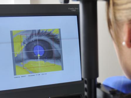
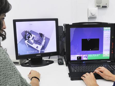
The BCBL is equipped with a Spectralis OCT device, which integrates all the features and tools of Spectralis. The integration of Heidelberg Engineering equipment allows us to perform a comprehensive analysis of the structural composition of the Optic Nerve Head (ONH) or the Nerve Fiber Layer (RNFL) by connecting to the HEYEXTM software platform.
The Spectralis system provides an excellent scan rate (40,000 A-scans/second) and is especially suitable for real-time imaging collection. It has an axial resolution (in tissue) of up to 3.9 μm (digital) and transverse resolution (in tissue) of 14 μm. A maximum field of view (30°x30°) can be scanned by the system. The isotropic high resolution mode is 5 μm. The device is also equipped with a high-resolution 24” Monitor (1920x1200), and a computer with Quad Core Processor. This system uses a remote desktop mount, thus minimizing eye motions.
The OCT in our center also includes available software capable of segmenting retinal layers beyond the RNFL. It is sufficient to automatically define ten boundaries within the retina and allows identification of up to 11 retinal layers (ILM, RNFL, GCL, IPL, INL, OPL, ONL, ELM, PR1, PR2, RPE). Our system can also be used to perform OCT Angiography to visualize blood vessels without the need for contrast agents.

